

What is digital imaging?
Digital imaging is a dental procedure that captures images of the structures, such as teeth and gums, inside a person’s mouth. Dental radiographs, more commonly referred to as X-rays, are the most commonly used form of digital imaging. Dental radiographs are used to diagnose various oral conditions and diseases and are essential in creating effective treatment plans for patients, identifying issues before they become major concerns. Digital images are both intraoral (inside the mouth) and extraoral (outside of the mouth).
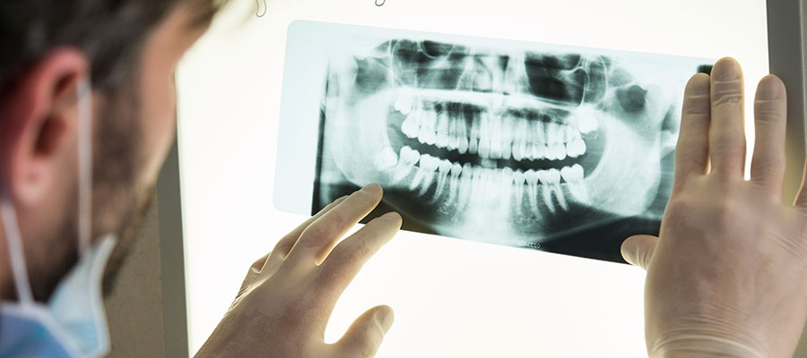
What are the different types of radiographs used by dentists?
Dentists use a variety of radiographs to diagnose, treat and prevent oral conditions including:
- Bitewing: visual of lower and upper posterior teeth
- Periapical: visual of the entire tooth
- Panoramic: visual of teeth, jaws, sinuses and nasal area
- Occlusal: visual of the upper and lower jaw, as well as the floor of the mouth.
The type of radiograph used depends on the dentist’s purposes. Dentists opt to use panoramic radiographs to help determine if orthodontic or implant placement is needed. Occlusal radiographs are excellent at allowing dentists to monitor the development of teeth, primarily in children. Bitewing radiographs help dentists to locate areas of decay in teeth, as well as to identify gum disease.
Why do dentists use digital imaging?
Digital imaging helps to achieve the following purposes for dentists:
- Find cavities and evidence of tooth decay
- Identify if teeth are impacted
- Examine the roots of teeth
- Examine the spacing between teeth
- Identify any damage within the tooth
- Identify the presence of periodontal or gum disease
- Monitor the development and growth of new teeth
- Monitor overall oral health as a preventative measure
- Locate cysts and tumors
- Identify oral infections
- Plan treatment for orthodontics or implants
Dentists depend on digital imaging as an important diagnostic tool to ensure the optimal oral care for their patients. Dental radiographs allow dentists to see the details of a patient’s teeth and the supporting tissues of the mouth that cannot be seen with the naked eye.